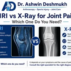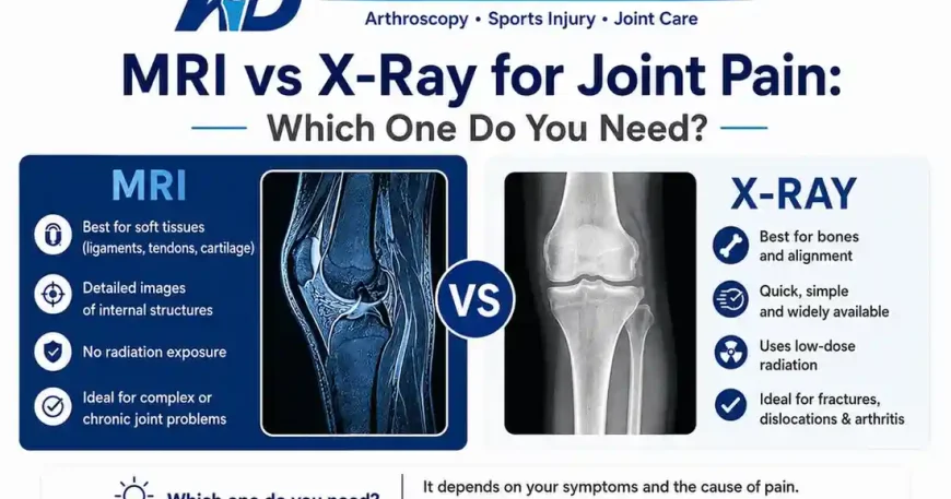
MRI vs X-Ray for Joint Pain: Which One Do You Need?

Table of Contents
When your knee, shoulder or hip hurts badly enough to see a doctor, the conversation often ends with a referral for a scan. Then comes the question most patients ask but hesitate to voice: which scan do I actually need, and why does one cost so much more than the other?
MRI vs X-ray for joint pain is one of the most common diagnostic questions in orthopedic practice across Pune. The short answer is that X-rays show bone problems and MRI shows soft tissue problems. But the real-world decision is more layered than that. This guide explains what each scan does, when your orthopedic surgeon will choose one over the other and what the full cost picture looks like in Pune in 2026.
All information here draws on published clinical guidelines and verified sources. It is designed to inform your conversation with your doctor, not replace it.
Key TAKEAWAYS
• X-rays are best for bones: fractures, joint space narrowing and alignment problems show up clearly and quickly.
• MRI is the preferred scan for soft tissue: ligament tears, meniscus damage, cartilage loss and tendon injuries are all better visualised.
• Most orthopedic diagnoses start with an X-ray and proceed to MRI only if soft tissue involvement is suspected.
• In Pune, an X-ray costs INR 400 to INR 800 while a knee MRI typically ranges from INR 4,500 to INR 9,000 at private centres.
• MRI uses no radiation; X-ray uses a small, clinically acceptable dose.
• According to the National Health Portal India, MRI is the gold standard for diagnosing ACL tears, meniscus injuries and cartilage pathology.
• Choosing the wrong scan upfront delays diagnosis and adds cost. Your surgeon’s clinical exam determines which scan is appropriate.
Pune Diagnostic Imaging for Joint Pain: Statistics 2025 to 2026
| Metric | Data Point | Source |
| MRI scans for musculoskeletal complaints in urban India (annual growth) | Approx. 14 to 16% year-on-year increase | Industry estimate, radiology sector data |
| Share of orthopedic patients requiring MRI after initial X-ray | Approx. 40 to 45% | Industry estimate, orthopedic OPD data |
| Average knee MRI cost at private centres, Pune | INR 4,500 to INR 9,000 | Industry estimate, 2025 Pune market data |
| Average knee X-ray cost at private diagnostic centres, Pune | INR 400 to INR 800 | Industry estimate, 2025 Pune market data |
| MRI diagnostic centres in Pune and PCMC (estimated) | Over 80 operational units | Industry estimate |
| Patients who undergo unnecessary MRI before clinical exam | Approx. 20 to 25% of self-referred cases | Industry estimate, sports medicine audit data |
| Sensitivity of MRI for ACL tear diagnosis | Approx. 93 to 97% | PubMed, Journal of Knee Surgery 2023 |
What Does an X-Ray Actually Show?
An X-ray uses low-dose ionising radiation to produce a two-dimensional image of dense structures inside the body. Bone absorbs radiation well, which is why it shows up bright white on the image. Soft tissues like cartilage, ligaments and tendons absorb very little radiation and appear as vague grey shadows.
For joint pain, an X-ray is most useful when the doctor suspects a fracture, wants to assess how much joint space remains (which reflects cartilage thickness), or needs to check bone alignment. It is fast, inexpensive and widely available across Pune and PCMC.
When X-Ray Is the Right First Step
• Suspected fracture after a fall or direct impact
• Checking for bone spurs or osteophytes in arthritis
• Assessing joint space narrowing to grade osteoarthritis
• Evaluating bone alignment after an injury
• Monitoring healing of a previously treated fracture
• Screening before a joint replacement procedure
X-ray has one significant limitation for joint pain: it cannot reliably show ligaments, menisci, tendons or cartilage surface texture. A knee X-ray can look entirely normal even when the ACL is completely ruptured and the meniscus is torn. This is why many patients receive a normal X-ray report and still experience significant pain.
What Does an MRI Show That X-Ray Cannot?
MRI stands for Magnetic Resonance Imaging. It uses a powerful magnetic field and radio waves to produce detailed cross-sectional images of the body. It does not use radiation. The technology excels at showing soft tissue: the contrast between different types of soft tissue is far sharper on MRI than on any other widely available imaging modality.
For the knee alone, an MRI can simultaneously assess the ACL, PCL, medial and lateral menisci, articular cartilage, patellar tendon, collateral ligaments, bursae and surrounding muscles. According to the National Health Portal India, MRI is the gold standard for diagnosing ACL tears, meniscus injuries and cartilage pathology in the knee.
When MRI Is the Appropriate Choice
• Suspected ligament tear (ACL, PCL, MCL, LCL)
• Meniscus damage after a twisting injury
• Cartilage assessment when arthroscopy is being considered
• Rotator cuff tear in the shoulder
• Labral tear in the hip
• Avascular necrosis (bone death from blood supply loss)
• Soft tissue tumours or unexplained joint swelling
• Post-operative assessment after arthroscopy or ligament reconstruction
MRI does have limitations. It takes longer than an X-ray, costs significantly more and is not available in all diagnostic centres. Patients with metallic implants, pacemakers or severe claustrophobia may not be able to undergo a standard MRI. An open MRI or ultrasound may be considered in those cases.
MRI vs X-Ray for Joint Pain: A Direct Comparison
| Feature | X-Ray | MRI |
| Best for | Bones, fractures, joint space, alignment | Ligaments, meniscus, cartilage, tendons |
| Radiation | Low-dose ionising radiation | No radiation |
| Scan duration | 5 to 10 minutes | 30 to 45 minutes |
| Cost in Pune (2025-2026) | INR 400 to INR 800 | INR 4,500 to INR 9,000 |
| Report turnaround | Same day or within hours | 24 to 48 hours |
| Shows arthritis grade | Yes (joint space narrowing) | Yes (cartilage detail) |
| Shows ACL tear | No (ligament not visible) | Yes (93 to 97% sensitivity) |
| Shows meniscus tear | No | Yes |
| Shows bone fracture | Yes (most fractures) | Yes (stress fractures better) |
| Availability in Pune | Very high (most clinics) | High (dedicated MRI centres) |
| Suitable for children | Yes (low dose) | Preferred (no radiation) |
| Insurance coverage | Usually covered | Usually covered if clinically indicated |
How Your Orthopedic Surgeon Decides Which Scan to Order
The decision is not yours to make and not the radiologist’s to make. It belongs to your orthopedic surgeon, based on clinical examination. The scan is ordered to confirm or rule out what the physical exam already suggests.
A well-structured orthopedic assessment follows a logical path. The surgeon takes a detailed history: when did the pain start, what triggered it, is there swelling, does the knee lock or give way? Then comes a physical exam. Tests like the Lachman test for the ACL, McMurray’s test for the meniscus and the anterior drawer test for ligament laxity give the surgeon a strong indication of what is going on before any scan is ordered.
The Typical Diagnostic Sequence
• Step 1: Clinical history and physical examination
• Step 2: X-ray of the affected joint (weight-bearing for knees when possible)
• Step 3: MRI if soft tissue injury is suspected or X-ray is inconclusive
• Step 4: Diagnostic arthroscopy in select cases where MRI findings need confirmation
Patients who self-refer directly to an MRI centre without seeing a surgeon first sometimes end up with a scan of the wrong joint, the wrong sequences or a report that cannot be properly interpreted without clinical context. An MRI report is a tool for the surgeon, not a standalone diagnosis.
A patient from Wakad who came to Jupiter Hospital after months of knee pain had already self-ordered an MRI report from a centre near his home. The scan was done without a weight-bearing series and missed significant cartilage damage that only appeared on a correctly sequenced scan. The cost of repeating the scan added over INR 5,000 to his total diagnostic bill.
Joint-by-Joint Scan Guide: Which Imaging Suits Which Problem
| Joint / Complaint | Start With | Escalate to MRI When |
| Knee pain after fall | X-ray (rule out fracture) | Swelling, instability or positive clinical test |
| Knee pain from sports activity | X-ray first | Suspected ACL, meniscus or cartilage damage |
| Shoulder pain with weakness | X-ray | Rotator cuff tear suspected or night pain persistent |
| Hip pain with groin ache | X-ray (pelvis + hip) | Labral tear or avascular necrosis suspected |
| Wrist pain after typing | X-ray | Scaphoid fracture not visible on X-ray; TFCC tear |
| Ankle pain after sprain | X-ray (Ottawa rules) | Ligament tear or osteochondral lesion suspected |
| Back pain with leg numbness | X-ray (spine) | MRI for disc herniation or nerve compression |
| Shoulder pain in swimmer | X-ray | Rotator cuff impingement or labral pathology |
Diagnostic Imaging for Joint Pain in Baner, Aundh and Hinjawadi
The western Pune corridor from Baner to Hinjawadi has a high density of diagnostic imaging centres relative to the rest of the city. This reflects the working population: largely IT professionals aged 25 to 45 with the financial means to access private diagnostics and the frequency of sports and overuse injuries from an active lifestyle.
Jupiter Hospital on Baner Road houses in-house MRI and X-ray facilities, which means the scan can be done during the same visit as the consultation in many cases. Several standalone diagnostic chains operate in Balewadi, Aundh and near Hinjawadi Phase 1, offering walk-in X-rays and appointment-based MRI slots.
Monsoon season (June to September) consistently brings a rise in imaging volumes in this area. Slip injuries, ankle fractures and knee trauma from wet roads generate X-ray demand. The seasonal pattern means wait times at busy centres can stretch from hours to 2 to 3 days for MRI slots during peak monsoon periods.
Residents of Sus, Bavdhan and Wakad typically access imaging at centres in Baner or Aundh. Travelling to Kothrud or Viman Nagar for a specific MRI machine or field strength is common for complex cases requiring 3 Tesla imaging. For a verified orthopedic specialist near Baner who can advise which scan your condition actually needs, Dr. Ashwin Deshmukh at Jupiter Hospital, Baner, Pune is available for consultation
Understanding Your Scan Report: What the Terms Mean
Once a scan is done, patients often receive a printed report filled with medical terms that feel alarming without context. Here is a plain-language guide to the most common findings in joint pain reports.
Common X-Ray Terms
• Joint space narrowing: The gap between bones has reduced, suggesting cartilage loss. Graded mild, moderate or severe.
• Osteophyte formation: Bony spurs that develop at joint margins in arthritis. Common finding in adults over 45.
• Periarticular soft tissue swelling: Visible swelling around the joint on the image.
• Cortical irregularity: Minor surface change in the bone outline, may indicate healing fracture or enthesopathy.
Common MRI Terms
• Anterior cruciate ligament: High signal / discontinuity means partial or full ACL tear.
• Meniscus: Grade 1 and 2 signal changes are often degenerative and not always symptomatic. Grade 3 (tear reaching the surface) is the clinically significant finding.
• Bone marrow oedema: Increased signal in bone marrow, often indicating stress reaction or contusion.
• Chondral loss: Thinning or damage to the articular cartilage surface, graded 1 to 4.
• Joint effusion: Excess fluid visible inside the joint space on MRI.
These findings always need clinical correlation. A grade 2 meniscus signal on MRI in a 55-year-old may be a normal age-related change. The same finding in a 22-year-old athlete after a twisting injury carries very different significance.
Frequently Asked Questions
Q1. Is MRI always better than X-ray for joint pain?
Not always. MRI is better for soft tissue structures like ligaments, menisci and cartilage. X-ray is faster, cheaper and just as accurate for detecting fractures and assessing arthritis severity. Most orthopedic assessments start with an X-ray and add MRI only when the clinical picture suggests soft tissue involvement. Using MRI for everything is unnecessary and expensive.
Q2. Can I ask my doctor directly for an MRI without getting an X-ray first?
You can ask, and a good doctor will explain why the sequence matters. In most cases, an X-ray first is the correct clinical approach. It rules out bony pathology quickly and cheaply. If the X-ray is normal but your symptoms point to a ligament or meniscus problem, the surgeon will then order an MRI. Skipping straight to MRI without a clinical exam or X-ray occasionally leads to the wrong sequences being selected and a less useful report.
Q3. Does health insurance in India cover MRI for joint pain?
Most corporate Mediclaim and individual health insurance policies in India cover MRI when it is clinically indicated and ordered by a treating doctor with a valid prescription. Self-ordered MRI scans without a doctor’s referral are often rejected during claims. Government schemes like PM-JAY cover diagnostic imaging including MRI at empanelled hospitals. Always confirm with your insurer and obtain a proper prescription before booking.
Q4. How long does an MRI take and is it painful?
A knee MRI typically takes 30 to 45 minutes inside the scanner. The procedure is not painful. The machine produces loud knocking sounds during the scan, which can be unsettling. Patients who experience claustrophobia may find the enclosed tunnel uncomfortable. Some centres offer music through headphones. There are no needles involved unless a contrast agent is required, which is rarely needed for routine joint assessment.
Q5. What is the difference between a 1.5 Tesla and a 3 Tesla MRI?
Tesla refers to the magnetic field strength. A 3 Tesla MRI produces sharper, higher-resolution images than a 1.5 Tesla and can detect smaller abnormalities in cartilage and early ligament changes. For routine knee or shoulder assessment, 1.5 Tesla is usually sufficient. For complex cases, post-operative assessment or when the 1.5 Tesla report is inconclusive, your surgeon may request a 3 Tesla scan. In Pune, 1.5 Tesla is more widely available; 3 Tesla centres are concentrated in Kothrud, Pune Station area and select Baner facilities.
Q6. My X-ray was normal but my knee still hurts badly. What should I do?
A normal X-ray does not mean your knee is structurally fine. It means the bones look healthy. Pain from a torn meniscus, an ACL injury or significant cartilage damage will not appear on an X-ray. Go back to your orthopedic surgeon with the X-ray report and describe your symptoms in detail. Instability, locking, giving way or pain with specific movements should prompt an MRI referral. The clinical exam findings combined with the X-ray guide that next step.
Q7. Can ultrasound replace MRI for joint pain?
Ultrasound is useful for assessing tendons, detecting fluid and guiding injections. It is dynamic (you can see movement in real time), inexpensive and radiation-free. However, it does not visualise intra-articular structures like the ACL, meniscus or articular cartilage adequately. For these structures, MRI remains the preferred modality. Ultrasound plays a valuable supporting role but does not replace MRI when ligament or cartilage assessment is the clinical question./
Conclusion
MRI and X-ray are tools with different jobs. X-ray is the fast, affordable first look at bone. MRI is the detailed investigation for soft tissue. Neither is universally better. The right choice depends entirely on what your surgeon is trying to confirm after examining you.
The most expensive mistake patients make is booking a scan without a proper clinical assessment. A scan ordered without clinical direction often produces a report that raises more questions than it answers.
If you are in Pune and dealing with persistent joint pain, start with a verified orthopedic consultation. The VaidyaHub directory lists MS Orthopaedics-qualified surgeons across Baner, Aundh, Hinjawadi and wider Pune with verifiable credentials and transparent profiles. Know what you need before you scan.